Arachnoid cysts of the posterior fossa are rare. Superiorly the cerebellum is separated from the cerebral hemispheres by the tentorium cerebelli.
Getting To The Bottom Of A Medulloblastoma Mystery St Jude Progress
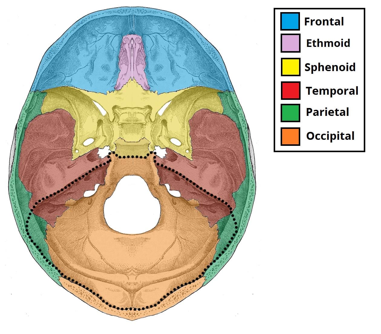
It is said to be butterfly shaped with a middle part accommodating the pituitary gland and two lateral parts accommodating the.
. The middle cranial fossa is located as its name suggests centrally in the cranial floor. The right paraduodenal fossa fossa of Waldyer the foramen of Winslow lesser sac a hole in the mesentery transmesenteric a hole in the transverse mesocolon. The inferior ileocecal fossa.
The portion above the spine is called the supraspinous fossa and that below it the infraspinous fossa. The posterior fossa accommodates the cerebellum and brain stem. Each fossa accommodates a different part of the brain.
The brain stem and cerebellum. It is bounded in front by the posterior margins of the lesser wings of the sphenoid bone the anterior clinoid processes and the ridge forming the anterior margin of the. Its fibres combine with the tendon of the psoas major.
The two fossae are connected by the spinoglenoid notch situated lateral to the root of the spine. Hover onoff image to showhide findings. When arachnoid cysts are encountered the presenting symptoms are frequently otologic with hearing loss and imbalance occurring commonly.
They are known as the anterior cranial fossa middle cranial fossa and posterior cranial fossa. The middle cranial fossa deeper than the anterior cranial fossa is narrow medially and widens laterally to the sides of the skullIt is separated from the posterior fossa by the clivus and the petrous crest. Three cases are presented with a previously unreported otologic symptom that of bilateral hearing loss which in one case was fluctuant.
The superior ileocecal fossa. Click image to align with top of page. None of the patients had the common.
Originates from surface of the iliac fossa and anterior inferior iliac spine. M1 Anterior Neck and Thorax Back and Spinal Cord Larynx Pharynx and Cervical Sympathetic Trunk Superior Mediastinum and Root of Neck Heart Lungs and Ventilation Pathway Posterior Mediastinum Kidneys and Urinary System Abdominal Wall Peritoneum and Intestines Stomach Liver and Spleen Duodenum. The left paraduodenal fossa fossa of Landzert.
Tap onoff image to showhide findings. The back of the scapula also called the dorsal or posterior surface is arched from above downward and is subdivided into two unequal parts by the spine of the scapula. Posterior fossa - axial CT image at level of pons.
It combines with the psoas major to form the iliopsoas the major flexor of the thigh. The iliacus muscle is a fan-shaped muscle that is situated inferiorly on the posterior abdominal wall.
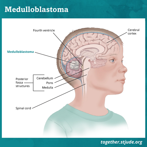
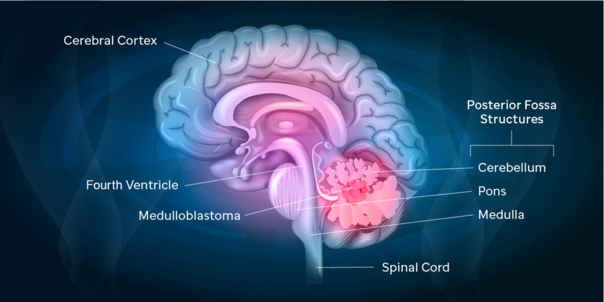
Posterior Fossa Syndrome Together
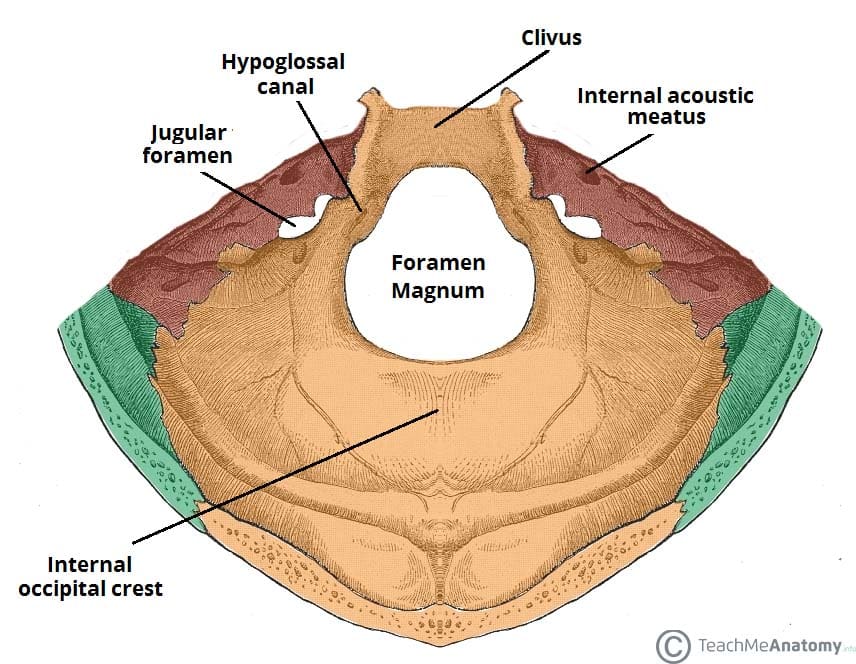
Posterior Cranial Fossa Boundaries Contents Teachmeanatomy

Posterior Cranial Fossa Wikipedia

Posterior Cranial Fossa Wikipedia
Posterior Cranial Fossa Boundaries Contents Teachmeanatomy

Figure Anatomy Of The Brain The Pdq Cancer Information Summaries Ncbi Bookshelf
Brain Tumors In Children Weill Cornell Brain And Spine Center

Posterior Cranial Fossa Radiology Reference Article Radiopaedia Org
0 comments
Post a Comment